Nerve cells
Nerve cells, or neurons, are specialized cells that form the basis of the nervous system. They have a unique structure and function that allows them to transmit electrical and chemical signals throughout the body.
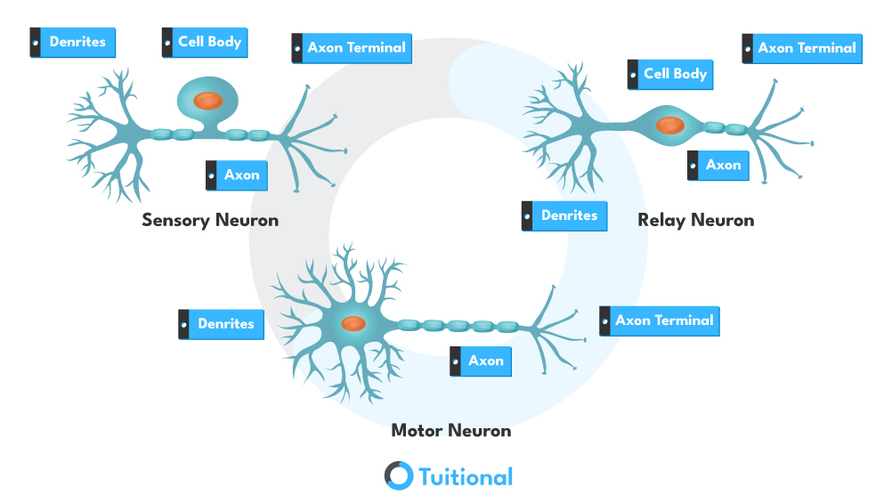
Sensory Neurons: Sensory neurons are responsible for transmitting impulses from sensory organs, such as the eyes, ears, skin, and internal organs, to the central nervous system (CNS). These impulses provide information about changes in our surroundings or within our bodies. Sensory neurons enable us to perceive sensations like sight, sound, touch, taste, and smell.
Motor Neurons: Motor neurons carry impulses from the CNS to effectors, such as muscles and glands, resulting in a response. These impulses initiate muscle contractions or glandular secretions, allowing for movement and physiological functions.
Interneurons (Relay Neurons): Interneurons, also known as relay neurons, are found exclusively within the CNS. They serve as connectors between sensory and motor neurons, facilitating communication and integration of signals within the nervous system. Relay neurons are neither sensory nor motor; instead, they relay information between different neurons inside the CNS. They play a crucial role in processing and integrating sensory information, as well as coordinating motor responses.
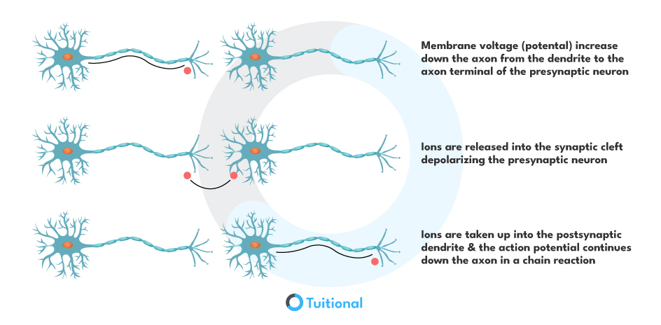
The junctions where neurons connect with each other are called synapses. At synapses, electrical impulses are converted into chemical signals (neurotransmitters), which then relay the signal from one neuron to the next. This allows for communication and transmission of information within the nervous system.
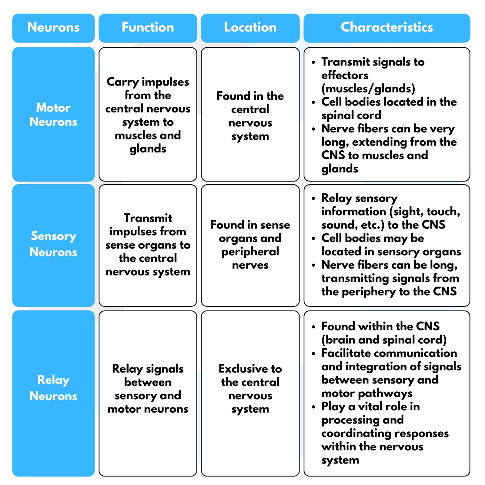
Types of Neurons:
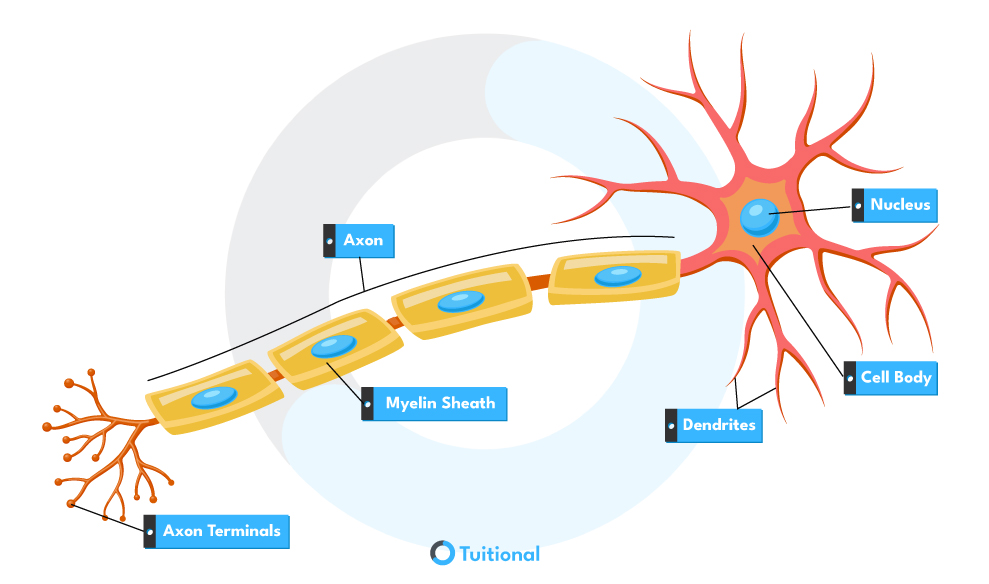
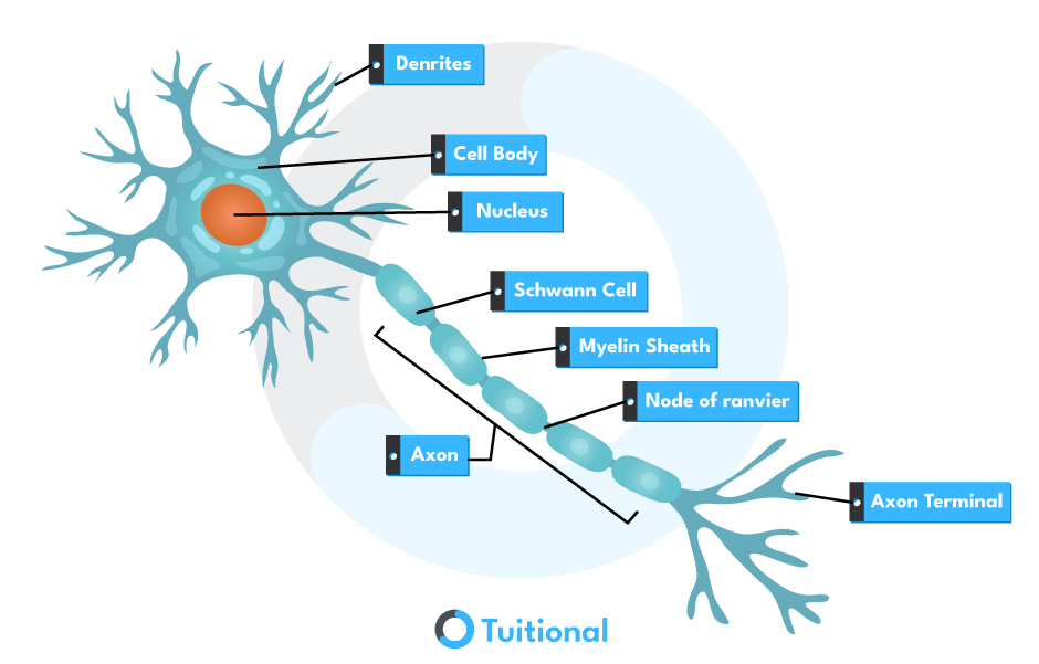
Structure of Neurons:
Neurons, the fundamental units of the nervous system, have a complex structure optimized for transmitting electrical impulses. At the core of each neuron lies the cell body, also known as the soma, which contains the nucleus and most of the organelles responsible for cellular functions. The cytoplasm fills the cell body, facilitating biochemical processes necessary for the neuron’s survival and function. It’s within the cell body that the decision to generate an electrical signal, called an action potential, is made based on the inputs received from other neurons through its dendrites.
Extending outward from the cell body are numerous branching structures known as dendrites. These dendrites serve as the primary sites for receiving signals from other neurons. They contain receptors that can detect neurotransmitters released by neighboring neurons. The dendrites collect and integrate these signals, summing them up to determine whether to generate an action potential. Once a threshold is reached, an action potential is initiated and travels along the neuron’s axon.
The axon is a long, slender projection extending from the cell body that carries the action potential away from the cell body toward other neurons or effector cells. Surrounding the axon is the myelin sheath, a fatty substance produced by glial cells that acts as an insulating layer. This myelin sheath facilitates the rapid transmission of electrical impulses by preventing signal leakage and allowing the action potential to “jump” from one node of Ranvier to the next in a process called saltatory conduction. Ultimately, the action potential reaches the nerve terminals, where it triggers the release of neurotransmitters into the synapse, allowing communication with the next neuron or target cell in the circuit.
The directional flow of signals in sensory and motor fibres is crucial for the proper functioning of the nervous system, enabling sensory input to be processed centrally and appropriate motor responses to be initiated peripherally. This uni-directional flow ensures efficient sensory perception and motor control, allowing organisms to interact with their environment and respond to stimuli effectively.
Action Potential:
An action potential is a rapid change in the voltage across a membrane. The membrane potential is determined by the relative ratio of ions in the extracellular space compared to the intracellular plus the permeability of each ion.